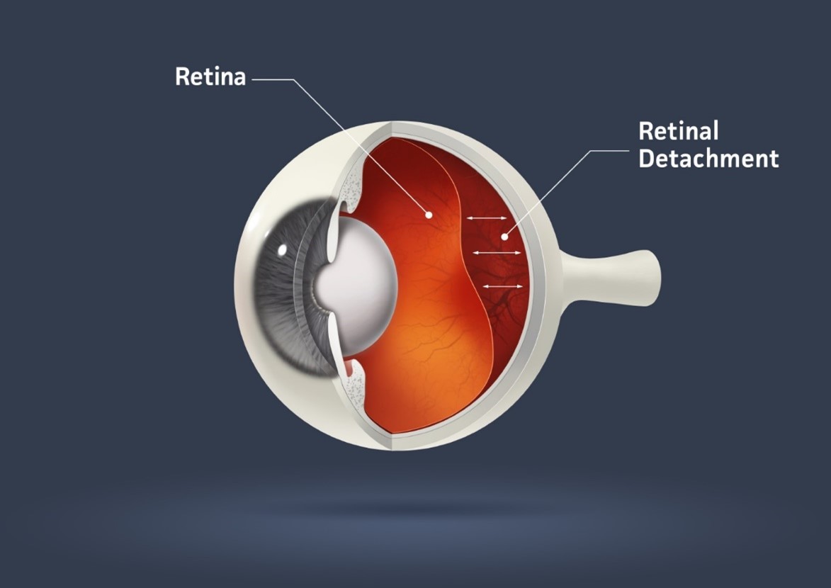
Retinal detachment is a serious eye condition that requires immediate medical attention to prevent permanent vision loss. The retina is the thin layer of tissue at the back of the eye that converts light into nerve signals, which are then sent to the brain to form images. When the retina detaches from its underlying tissue, it can lead to severe vision impairment or blindness if not treated promptly.
What is Retinal Detachment?
Retinal detachment occurs when the retina becomes separated from the back of the eye. The retina is essential for vision, as it receives light and sends visual information to the brain. When it detaches, the retina can no longer function properly, and it can result in permanent loss of vision in the affected eye.
There are three types:
- Rhegmatogenous Retinal Detachment: This is the most common type and occurs when a tear or hole in the retina allows fluid to leak under the retina, causing it to detach.
- Exudative Retinal Detachment: This type occurs when fluid builds up beneath the retina without a tear or hole, often due to underlying conditions like inflammation or vascular diseases.
- Tractional Retinal Detachment: This occurs when scar tissue, often caused by diabetic retinopathy, pulls on the retina, causing it to detach.
Causes of Retinal Detachment
There are several risk factors and causes of retinal detachment. Some of the most common include:
- Aging: As you age, the vitreous gel inside the eye shrinks and may pull away from the retina, increasing the risk of retinal tears or detachment.
- Trauma or Injury: A severe blow to the eye or head can cause retinal detachment, especially if it leads to a tear in the retina.
- High Myopia (Nearsightedness): People with severe nearsightedness are at a higher risk due to the elongated shape of their eyes, which can put additional stress on the retina.
- Previous Eye Surgery: Surgeries like cataract removal can increase the risk of retinal detachment.
- Family History: If a close family member has had retinal detachment, your risk of developing the condition may be higher.
- Other Eye Diseases: Conditions like diabetic retinopathy, eye infections, or inflammation can cause retinal detachment.
Symptoms of Retinal Detachment
Recognizing the symptoms of retinal detachment early is crucial for preventing permanent vision loss. Common symptoms include:
- Sudden Appearance of Floaters: These are dark, shadowy spots or lines that appear in your field of vision. They may seem to float or drift around as you move your eyes.
- Flashes of Light: You may experience sudden flashes of light, especially in the peripheral vision, which can signal the retina is being pulled.
- Blurry or Distorted Vision: The vision in the affected eye may become blurry or distorted, with some parts of the visual field appearing missing or shadowed.
- Loss of Peripheral Vision: A person may notice that their side vision (peripheral vision) is gradually lost, leaving only central vision.
- A Curtain Over Your Vision: This is a common symptom of a retinal detachment where part of the vision appears as if a dark curtain has been drawn across the field of vision.
If you experience any of these symptoms, it’s important to see an eye doctor immediately. The earlier retinal detachment is detected, the better the chances of preserving vision.
Diagnosis of Retinal Detachment
To diagnose retinal detachment, an ophthalmologist will perform a comprehensive eye exam, which may include:
- Dilated Eye Exam: The doctor will use eye drops to dilate your pupils, allowing them to examine the retina more thoroughly.
- Ophthalmoscopy: This procedure uses a special light and magnifying lens to closely examine the retina.
- Ultrasound: If the retina is obscured by bleeding or other obstructions, an ultrasound may be performed to get a clearer view of the retina.
- Optical Coherence Tomography (OCT): This imaging test creates detailed cross-sectional images of the retina, allowing the doctor to identify areas of detachment.
Treatment of Retinal Detachment
The treatment for retinal detachment depends on the type, severity, and location of the detachment. Early intervention is crucial to prevent permanent vision loss. Some common treatment options include:
- Laser Surgery (Photocoagulation): If there are small retinal tears or holes, a laser can be used to seal the tears and prevent further detachment. This method is often used in cases of rhegmatogenous detachment.
- Cryopexy (Freezing Treatment): Cryopexy is another technique used to treat retinal tears. In this procedure, a freezing probe is applied to the outside of the eye to create scar tissue, which helps seal the tear and prevent fluid from getting underneath the retina.
- Pneumatic Retinopexy: This treatment involves injecting a small bubble of gas into the eye to push the retina back into place. Laser treatment is then used to seal the tear, and the gas bubble gradually resorbs over time.
- Scleral Buckling: In cases of more severe detachment, a small band (scleral buckle) is placed around the eye to help reattach the retina. This procedure helps to relieve the tension pulling on the retina.
- Vitrectomy: If the detachment is due to scar tissue or if other methods are unsuccessful, a vitrectomy may be performed. This surgery involves removing the vitreous gel from the eye and replacing it with a gas bubble or silicone oil to push the retina back into place.
Recovery and Outlook
The recovery process after retinal detachment surgery varies depending on the treatment used and the severity of the detachment. Patients may need to avoid certain activities, like heavy lifting or strenuous exercise, for several weeks. In some cases, the patient may need to keep their head in a certain position to help ensure the retina stays in place during healing.
Vision recovery can be gradual, and it may take several weeks or even months for the full effects of surgery to be seen. However, if retinal detachment is caught early and treated promptly, there is a good chance of restoring vision.
Preventing Retinal Detachment
While retinal detachment cannot always be prevented, there are steps you can take to reduce your risk:
- Regular Eye Exams: If you have risk factors like high myopia or a family history of retinal problems, schedule regular eye exams to catch issues early.
- Manage Health Conditions: Controlling conditions like diabetes and high blood pressure can help prevent complications that could lead to retinal problems.
- Protect Your Eyes: Wearing protective eyewear during high-risk activities or sports can prevent trauma to the eyes.
Retinal detachment is a serious eye condition that can lead to permanent vision loss if not treated promptly. Understanding the causes, symptoms, diagnosis, and treatment options for retinal detachment is essential for protecting your vision. If you experience any symptoms like floaters, flashes of light, or a curtain-like shadow in your vision, seek medical attention immediately.
Don’t wait for symptoms to worsen—schedule an eye exam today to at Paranjpe Eye Clinic protect your vision and ensure your eyes stay healthy for years to come!

